Ptychography – With a “P” As in “Pterodactyl”
A 20-minute read
Ptychography is an area-by-area scanning technique in which a constant, coherent illumination is scanned across a region of a sample in many adjacent and overlapping positions. The scattered radiation is allowed to propagate, and the resulting diffraction pattern is measured using a 2D, pixelated detector. The redundancy in this dataset is then used, together with an iterative optimization method, to create quantitative, full-field images with diffraction-limited spatial resolution and sensitivity to both amplitude and phase contrast.
But how does Ptychography work? And, perhaps most importantly, is it worth all the hype?
With short wavelengths (of a few picometers), electron microscopes are capable of resolving individual atoms within a lattice. Thanks to recent breakthroughs in electron detector technology, and the adoption of innovative imaging methods, the main factors that affect performance have shifted from instrument stability and aberrations to sample stability and resilience to a dose [1].
The key breakthrough driving this shift is a technique that can perform full-field, quantitative imaging of arbitrary samples, largely independent of the shape and quality of the illumination. It is extendable to partially coherent illumination, it is capable of spectrally-resolved and 3D imaging – and it lifts the need for image-forming optics from the system. This technique is called “Ptychographic Coherent Diffractive Imaging” (CDI), or simply “Ptychography”, and it has sparked a revolution in imaging science.

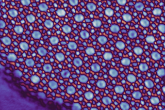
Top: A working principle of a ptychographic scan performed on a simulated 20-nm Nickel Siemens Star, which is deposited on a 200-nm Silicon membrane. The illumination function is focused on the sample, and 2D diffraction patterns are collected from partially overlapping regions in an area-by-area scan.
Bottom: Information obtained from the ptychographic reconstruction: amplitude (left) and phase (right), which are separated from the full-field profile of the probing illumination (inset).
The Ptychography Imaging Problem
Modern Ptychography is a phase retrieval technique with roots in Hoppe’s work on Crystallography in the 1960s [2]. The current implementation of this technique uses many adjacent, overlapping scattering measurements to recover the sample using iterative, non-linear inversion techniques. Usually, these measurements consist of a series of 2D diffraction patterns, which are obtained by moving a finite-area probe over a region of interest in a sample.
Key criteria relating to this approach are: (i) the overlap between the scanning positions, which is usually between 60% and 80%; and (ii) an upper limit on the beam’s diameter, to ensure proper sampling of the diffraction during the measurement. Interestingly, there are no further constraints on the sample’s geometry or composition or beam profile. At the end of the phase retrieval process, the effects of the sample and probing beam are disentangled from each other, yielding two complex-valued images. The first is a quantitative, complex transmission—or reflection map—of the sample, and the other is a full-field profile of the probing illumination.
The complex nature of the sample map means that Ptychography provides quantitative information about the amplitude and phase contrasts of a sample. The amplitude contrast shows the sample’s projected material composition, while the phase contrast yields both material and topographic information. For thin samples, such as 2D materials, these images can produce a map of the sample’s composition and morphology, with performance exceeding that of the image-forming optics that are used in TEMs [3]. In addition, the wavefront image provides feedback on the performance of the microscope’s beam delivery system, and it can be used directly to characterize the aberrations in the imaging system.
Further work has relaxed Ptychography’s imaging requirements by incorporating more complex image formation models into the reconstruction process. These include multiple, mutually incoherent beam or object modes that account for apparent points of incoherence in the recorded data, such as fluctuations in the beam, vibrations of the sample, or a broad spectrum in the scanning illumination [4, 5].
Also, multi-slice propagation methods can be used in the forward model of the problem to account for multiple scattering effects due to a thick sample. This method recovers a 3D structure by simultaneously solving for the multiple planes within a sample and expanding Ptychography’s imaging capabilities to 3D [6].
Ptychography’s Phase Retrieval Algorithms
The rapid rise of Ptychography came with the advent of the first ptychographic phase retrieval algorithms. Broadly speaking, these algorithms seek to find an unknown object by iteratively enforcing constraints that are known about the object in reciprocal spaces. These might include the plane of the object (“sample space”) and its 2D Fourier transform, which is defined as the square root of the scatter pattern that is measured during an experiment (“detector space”).
These methods have been a subject of study since the 1970s with the creation of the well-known Gerchberg-Saxton algorithm [7], but the game-changer in Ptychography was the shift to a partially overlapping, area-scanning modality that provides a powerful constraint in sample space. Essentially, regions of an object that are illuminated by several different, but overlapping, scanning positions must be consistent. The object update benefits from the refinement that is provided by adjacent position updates, and this leads to faster algorithmic convergence and increased robustness to noise.
Once the phase retrieval routine has finished, two complex-valued images are returned. The first is a full-field (amplitude + phase) image of the complex object transmission, or a reflection map, depending on the imaging geometry. The second is a complex profile of the illumination, which is used to record the data. Since Ptychographic CDI is sensitive to both phase and amplitude contrasts, the object image can be converted to a 2D+1 map, which contains information about the sample’s material composition and thickness/surface profile. On the other hand, the full-field image of the probe allows for an in-depth analysis of the optical system’s performance. For example, a projection of the complex beam profile onto the Zernike polynomials allows for a quantitative assessment of the aberrations in the beam.
The Role of the Detector
Any Ptychographic CDI microscopist will tell you that the detector is the beating heart of the experiment. The detector’s specifications determine the overall geometry and parameter space of all other components in the system. Above all else, the basic requirement is that the detector must be pixelated. This is to ensure that the unique structure of the 2D diffraction pattern is captured accurately for the phase retrieval inversion process.
In general, Ptychography detectors need to tackle two main challenges: (i) they must have a high sensitivity to all the intensities that are present in the measurement, and (ii) they must record a diffraction pattern quickly enough that a full dataset is not subject to mechanical instabilities or beam drift within the imaging system. Problem (i) is related to a detector’s dynamic range; the highest angle scatter should be measurable without saturating the central component of a diffraction measurement, which can mean sensitivity over many orders of magnitude in the signal’s strength. Problem (ii), on the other hand, is directly related to the beam source’s stability and can vary quite a bit, depending on the source of an imaging system.
A Ptychography dataset itself can vary in size and duration, depending on the desired field-of-view for the image, the scattering strength of the sample that is under investigation, and the source’s overall flux. The exact number of diffraction patterns that are needed for a full Ptychography dataset is still an open research question; typically, however, about 50 are needed without any a-priori knowledge of the experiment. With system stability in the range of minutes, this means that each diffraction pattern must be collected in a matter of seconds, from initial exposure to full readout of the chip. This also forces the detector to have high sensitivity to high intensities, as integrating for long times over a weak beam is not feasible.
Ptychography is growing more and more widespread as better detector technology, software, and algorithms become more available and sophisticated. So, buckle up; larger diffraction datasets will come pouring in!
Discover our newest hybrid-pixel direct electron detector
References and Acknowledgments
[1] Chen, Z., Jiang, Y., Shao, Y.T. et al. Electron ptychography achieves atomic-resolution limits set by lattice vibrations. Science 372, 826-831 (2021).
[2] Hoppe, W. Beugung im inhomogenen Primärstrahlwellenfeld. I. Prinzip einer Phasenmessung von Elektronenbeungungsinterferenzen. Acta Crystallogr. A 25, 495–501 (1969).
[3] Jiang, Y., Chen, Z., Han, Y. et al. Electron ptychography of 2D materials to deep sub-ångström resolution. Nature 559, 343–349 (2018).
[4] Thibault, P. & Menzel, A. Reconstructing state mixtures from diffraction measurements. Nature 494, 68–71 (2013).
[5] Batey, D. J., Claus, D. & Rodenburg, J.M. Information multiplexing in ptychography. Ultramicroscopy 138, 13–21 (2013).
[6] Maiden, A.M. & Rodenburg, J.M. An improved ptychographical phase retrieval algorithm for diffractive imaging. Ultramicroscopy 109, 1256–1262 (2009).
[7] Gerchberg, R.W. & Saxton, W.O. A practical algorithm for the determination of phase from image and diffraction plane pictures. Optik 35, 237 (1972).
Brief Author Bios
Giulia Fulvia Mancini
Giulia is an Associate Professor in the University of Pavia’s Department of Physics and the Head of Research in the Laboratory for Ultrafast X-ray and Electron Microscopy (LUXEM). She obtained her M.Sc. degree from the University of Pavia and her Ph.D. in Physical Chemistry from the Federal Polytechnical School of Lausanne (EPFL). In 2015, she moved to the United States as a Postdoctoral Fellow at JILA, University of Colorado-Boulder and NIST (USA). After holding a Team Leader position as a Senior Research Associate at SwissFEL (PSI) with the EPFL, she returned to Italy as the winner of an ERC Starting Grant and a Cariplo Foundation Grant.
Charles Schrepferman Bevis
Charles received a B.A. in Physics from the University of Colorado-Boulder in 2011 and worked in the lab of Prof. Steve Cundiff, studying nonlinear interactions between matter and ultrashort laser pulses. He then obtained his M.Sc. (2017) and Ph.D. (2020) in Physics from the University of Colorado at Boulder—Kapteyn-Murnane Group. In 2021, Charlie joined LUXEM as a Postdoctoral Fellow, having obtained the prestigious Marie Skłodowska-Curie Postdoctoral Fellowship (HORIZON-MSCA-2021-PF-01-01 Action) to carry out the DECIPHER project at LUXEM. There, he leverages his expertise in computational imaging system design and algorithm development to create novel, correlative approaches using both electron and X-ray imaging.